In a nutshell
Every cell you have came from an existing cell dividing. This subtopic is about how that happens.
In eukaryotes, cells that can still divide follow a cell cycle, and the dividing part is mitosis, which makes two genetically identical daughter cells. Prokaryotes divide by binary fission instead, and viruses do not divide at all.
Assumed knowledge: Cell structure, Nucleic acids and DNA replication.
Core content
The cell cycle
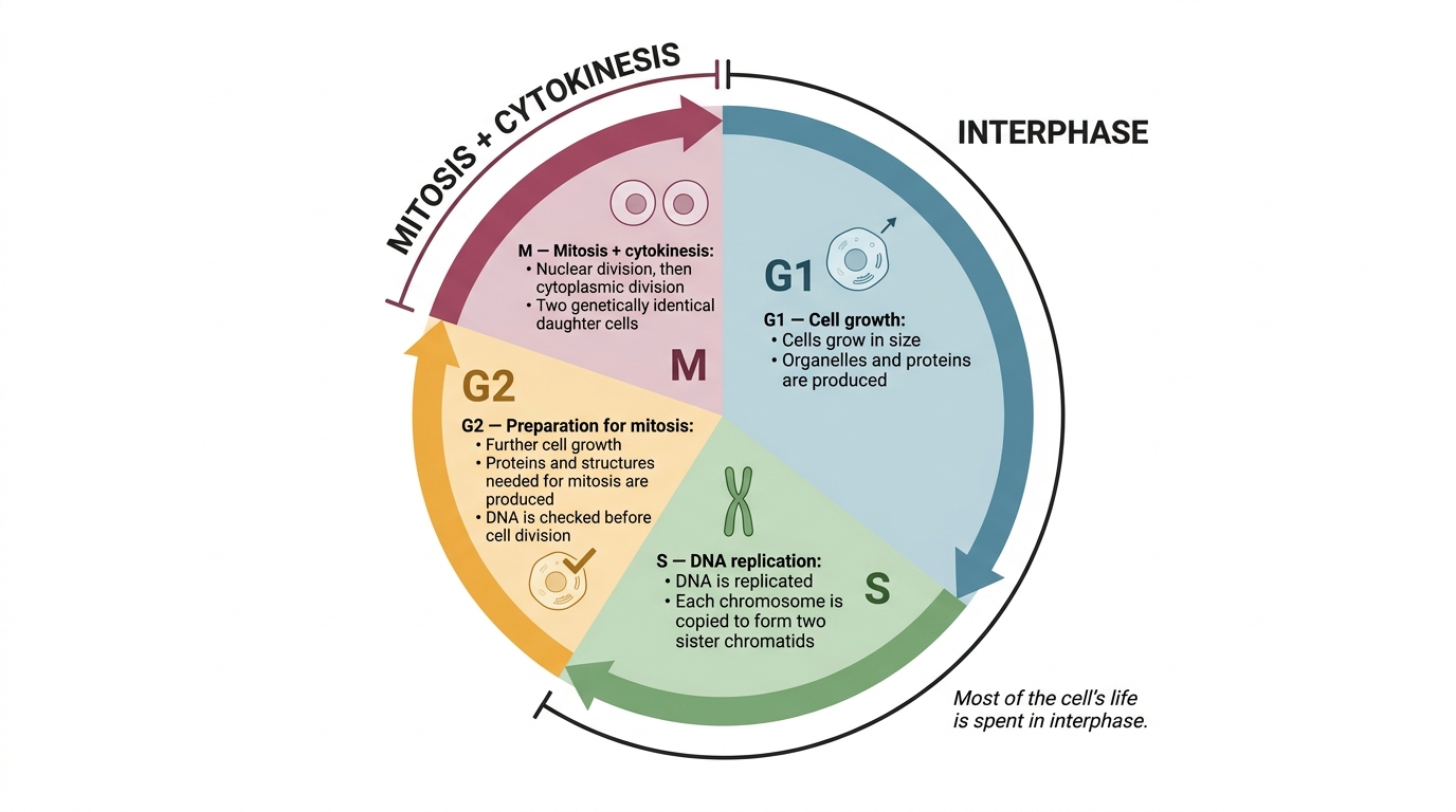
Within a multicellular organism, not all cells keep the ability to divide. The ones that do go through a repeating cell cycle with two parts:
- Interphase: the cell grows, replicates its organelles, and DNA replication takes place. This is the longest part of the cycle.
- Mitosis and cytokinesis: the nucleus divides (mitosis), then the cytoplasm divides (cytokinesis) to give two new cells.
During interphase, each chromosome is copied, so afterwards it consists of two identical sister chromatids joined at a centromere. The chromosomes are not condensed yet, so they are not visible under a light microscope.
DNA content through the cycle
The amount of DNA in the cell doubles during interphase (when DNA replicates in the S phase), stays doubled through mitosis, then halves as the cell splits in two.
The rising part is the S phase of interphase (DNA replication). The sudden drop is cytokinesis, where each daughter cell receives one full set.
The stages of mitosis
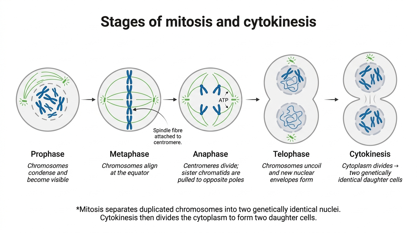
Mitosis is one continuous process, but it is described in four stages. Learn what the chromosomes are doing in each, because "explain the appearance" questions want the behaviour, not just the name.
| Stage | What the chromosomes do | Mark-winning detail |
|---|---|---|
| Prophase | Chromosomes condense and become visible, each as two sister chromatids joined at a centromere. The nuclear envelope breaks down and spindle fibres form. | "condensed / short and thick / visible" |
| Metaphase | Chromosomes line up along the equator (centre) of the cell. | Spindle fibres attach to the centromeres |
| Anaphase | The centromeres divide, and spindle fibres shorten to pull the sister chromatids to opposite poles. | Chromatids move in a V shape; this needs ATP |
| Telophase | Chromatids reach the poles, uncoil (become long and thin again), and a nuclear envelope reforms around each group. | Two new nuclei form |
Cytokinesis then divides the cytoplasm, giving two genetically identical daughter cells.
The spindle and the centromere
The spindle fibres are protein threads running from the poles of the cell. In metaphase they attach to the centromere of each chromosome; in anaphase they shorten and pull the sister chromatids apart to opposite poles. This is exactly what the centromere is for: it holds the sister chromatids together, attaches them to the spindle, and lets them be separated.
Chromosome, chromatid or homologous pair?
This is where most marks are lost, so get the words straight:
- A chromosome is one length of DNA. After replication it is two sister chromatids (identical copies) joined at the centromere.
- A homologous pair is two separate chromosomes carrying the same genes (but possibly different alleles), one inherited from each parent.
In mitosis, the spindle separates sister chromatids. Homologous chromosomes do not pair up and do not get separated: that is meiosis.
Still don't get it? · chromosome, chromatid or homologous pair?
Analogy: imagine a recipe book. One chapter is a chromosome. Just before the kitchen splits in two, someone photocopies every chapter and clips each copy to its original with a single paper clip. You now have one chapter made of two identical sheets, held together at the clip.
Rebuild it:
- The whole clipped thing is still one chromosome.
- Each identical sheet is a chromatid, and because they came from the same photocopy they are sister chromatids.
- The clip is the centromere, the single point holding the two sisters together.
- A homologous pair is a different idea: two separate chapters covering the same topics, one you got from your mother and one from your father. Same headings (genes), but the wording can differ (alleles).
Exam wording: after DNA replication, "each chromosome consists of two identical sister chromatids joined at the centromere". In mitosis the centromere divides and the sister chromatids separate. If you write that homologous chromosomes pair up, cross over, or separate, you have described meiosis, and the mark is gone.
Controlled and uncontrolled division: tumours and cancer
Mitosis is normally a tightly controlled process. If the controls fail, cells divide uncontrollably, and this can produce a tumour, and lead to cancer.
Because cancer is uncontrolled cell division, many cancer treatments aim to control the rate of cell division. Treatments such as chemotherapy and radiotherapy target rapidly dividing cells; a side effect is that they also harm the body's own fast-dividing healthy cells (for example those making blood cells or lining the gut).
Binary fission in prokaryotes
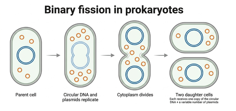
Prokaryotes (such as bacteria) do not use mitosis. They divide by binary fission:
- The circular DNA replicates, and the plasmids replicate too.
- The cytoplasm divides to produce two daughter cells.
- Each daughter cell gets one copy of the circular DNA and a variable number of plasmids.
There is no spindle and no mitosis.
Viruses do not divide
Viruses are non-living, so they cannot undergo cell division at all. Instead, a virus injects its nucleic acid into a host cell, and the host cell replicates the virus particles for it.
Required practical 2: root tip squash and the mitotic index
The practical prepares a stained squash of cells from a plant root tip, so the stages of mitosis can be seen and a mitotic index calculated.
Method (the reasons matter as much as the steps):
- Use a root tip, because it contains meristem tissue where cells are actively dividing by mitosis.
- Warm the tip in hydrochloric acid, to break down the tissue so the cells separate into a single layer.
- Add a stain (for example toluidine blue), because it binds to the DNA / chromosomes and makes them visible.
- Squash the tip gently under a coverslip, to spread the cells into a thin single layer so light can pass through.
The mitotic index is the proportion of cells that are in mitosis (chromosomes visible):
Give it as a decimal, unless the question asks for a percentage.
You should also be able to work out a cell's real size from a microscope image:
Worked examples
Model calculation: mitotic index and time in a stage.
A student counts the cells on a stained root tip slide and records the numbers below.
Total cells = 160 + 24 + 6 + 4 + 6 = 200. Cells in mitosis (all four mitotic stages) = 24 + 6 + 4 + 6 = 40.
Step 1, mitotic index:
Step 2, if one full cell cycle takes 20 hours, find the time spent in metaphase. The fraction of cells in a stage equals the fraction of the cycle spent in it:
Watch the units: 0.6 hours is 36 minutes, not "0 hours 6 minutes".
Model calculation: actual size from magnification.
A cell in a photomicrograph measures 30 mm across at a magnification of ×1500. Rearrange to make actual size the subject, and convert to a sensible unit:
Always put both lengths in the same unit before dividing.
Common exam mistakes
- Writing centriole when the answer is centromere. They are different structures, and "centriole" is rejected.
- Saying the spindle separates homologous chromosomes, or that chromosomes pair up or cross over, in mitosis. That is meiosis: in mitosis the spindle separates sister chromatids and homologous chromosomes never pair.
- Confusing a sister chromatid with a homologous chromosome, or writing that anaphase pulls "half a chromosome" apart. A whole chromatid moves to each pole.
- When asked to explain the appearance of a stage, only naming it. Naming earns nothing: describe what the chromosomes are doing (condensed and visible, lined up on the equator, being pulled to the poles).
- Confusing anaphase (chromatids being pulled apart) with telophase (chromatids arrived, nuclear envelope reforming).
- Claiming a tissue with more cells in interphase is dividing faster. It is the reverse: a higher proportion in mitosis (a higher mitotic index) means faster division.
- Spelling mitosis wrongly. It must be spelt correctly to earn the mark; examiners reject hybrids such as "meitosis".
- Calling prokaryotic division mitosis. Prokaryotes divide by binary fission, with no spindle.
- Giving the mitotic index as a percentage when a decimal is expected, and, in the magnification calculation, dividing without matching the units first or using the formula upside down.
Key definitions
- Mitosis - the part of the cell cycle in which a eukaryotic cell divides to produce two daughter cells, each with the identical copies of DNA produced by the parent cell during DNA replication.
- Cytokinesis - the division of the cytoplasm to produce two new cells.
- Centromere - the point holding the two sister chromatids together; it attaches them to the spindle and allows the chromatids to be separated to opposite poles.
- Mitotic index - the number of cells with visible chromosomes (in mitosis) divided by the total number of cells observed (usually given as a decimal).
- Binary fission - the division of a prokaryotic cell into two daughter cells, each with a single copy of the circular DNA and a variable number of copies of the plasmids.
- Actual size - the size of the image divided by the magnification.
Specification
- I can state that not all cells in a multicellular organism keep the ability to divide, and that those which do show a cell cycle.
- I can state that DNA replication occurs during interphase.
- I can define mitosis as producing two daughter cells with identical copies of the parent cell's DNA.
- I can describe the behaviour of the chromosomes in interphase, prophase, metaphase, anaphase and telophase, and recognise each stage.
- I can explain the appearance of the cells in each stage of mitosis.
- I can describe the role of spindle fibres attached to centromeres in separating the chromatids.
- I can state that division of the cytoplasm (cytokinesis) usually produces two new cells.
- I can explain that mitosis is controlled, that uncontrolled division can lead to tumours and cancers, and that many cancer treatments aim to control the rate of cell division.
- I can describe binary fission in prokaryotes: replication of the circular DNA and plasmids, then division of the cytoplasm into two cells, each with one copy of the circular DNA and a variable number of plasmids.
- I can state that viruses are non-living and do not divide, and that the host cell replicates the virus particles after their nucleic acid is injected.
- I can prepare and interpret a stained root tip squash, calculate a mitotic index, and calculate actual size using actual size = size of image / magnification (Required practical 2).
Related notes
Ready to test yourself?
Put All cells arise from other cells into practice with exam-style questions and full mark schemes.
Practise All cells arise from other cells